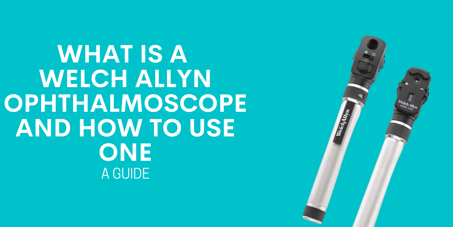
The Welch Allyn ophthalmoscope is an ophthalmoscope produced by revered medical technology producer Welch Allyn. It’s one of the label’s most popular products and is sold all over the world. It can be used as a complete diagnostic set or a standalone tool and it can also include an aneroid sphygmomanometer and otoscope.
If you’ve been looking for a quality ophthalmoscope, Welch Allyn Australia is the way to go. An ophthalmoscope is a diagnostic tool designed to examine the retina. If you have ever had your eyes tested or visited an ophthalmologist, there’s a good chance they used an ophthalmoscope to examine your retinas. There are two primary types of ophthalmoscope: indirect and direct. Indirect ophthalmoscopes check the entire retina while direct ophthalmoscopes are used to examine the retina’s centre.

Welch Allyn ophthalmoscopes utilise either SureColor LED technology or halogen illuminators. This allows medical professionals to see all elements of the retina as well as providing first class illumination.
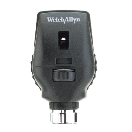
Welch Allyn ophthalmoscopes come in a variety of designs and sizes. The Welch Allyn pocketscope LED ophthalmoscope is known for being compact and easy to move around practices. The Welch Allyn 3.5 V ophthalmoscope is a more advanced version which contains a wealth of outstanding features, while the Welch Allyn Pocket Junior ophthalmoscope is the manufacturer’s most basic option.

Extra features of the Welch Allyn ophthalmoscope include a variety of dopter configurations, digital connectivity through the Welch Allyn iExaminer platform, rechargeable lithium-iron power handles for increased running time when up against other devices and advanced coaxial ophthalmoscopes designed to provide easy entry to the eye for enhanced field of view, reduced glare and true tissue colour

How do Welch Allyn ophthalmoscopes work?
Ophthalmoscopes work by illuminating either an undilated or dilated eye with a halogen or LED light. This provides the medical professional the opportunity to see the various aspects that make up the back of the eye to check for conditions or injuries. The part of that eye that ophthalmoscopes focus on is known as the “fundus”. It consists of the retina, a collection of blood vessels and the optic disc.
Ophthalmologists will examine the fundus when screening for conditions or injuries that affect the person’s eyes. It’s also standard practice to use an ophthalmoscope in eye examinations. An ophthalmoscope can examine the following:
● Optic nerve damage
● Retinal detachment or tear
● Glaucoma
● Macular degenerations
● Melanoma
● Diabetic retinopathy
● Infection
● Hypertension
The more advanced ophthalmoscope options provide medical specialists the
ability to alter the lens, aperture and aperture-lens combination to produce a greater view of the fundus. This can assist medical specialists in providing a more accurate diagnosis.
How to use Welch Allyn ophthalmoscopes
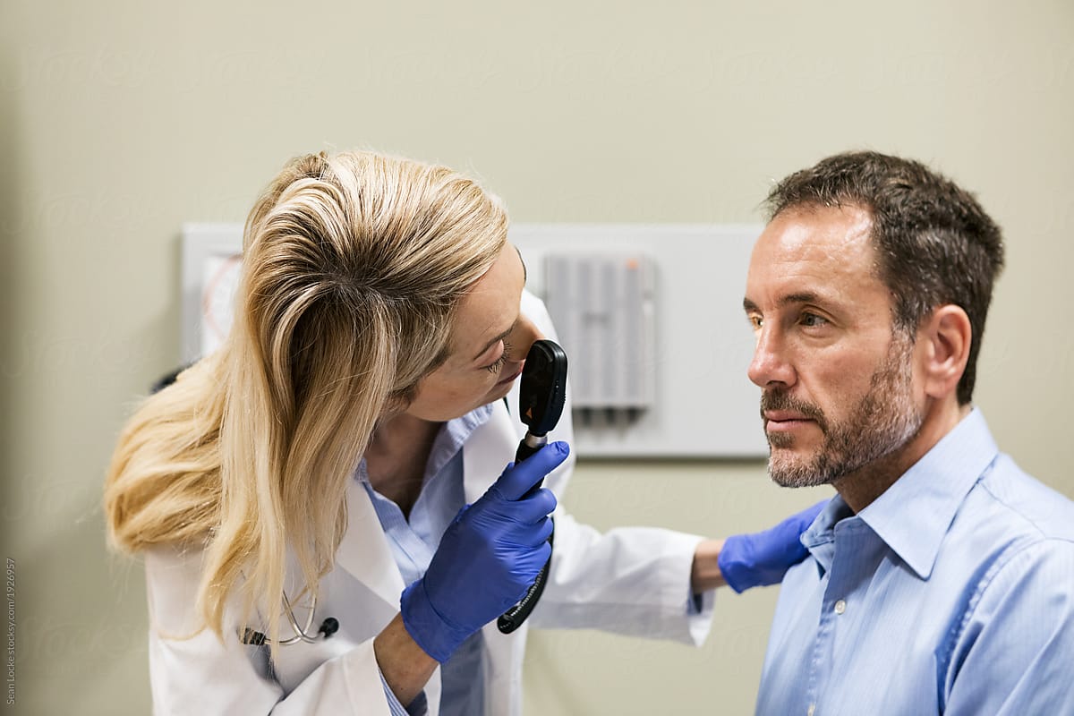
Ophthalmoscopes should always be operated by medical professionals. While the diagnostic equipment is non-invasive, improper use can still potentially cause eye damage.
When using the ophthalmoscope, it’s vital that the patient is still, seated and that the right working distance is maintained. Examination lights should be switched off completely, or turned down low, to optimise the view of the fundus.
Welch Allyn ophthalmoscopes are renowned for being highly intuitive. Adjustments can be made to the filter, lighting or lens simply by moving the dials and switches on the ophthalmoscope head. Most adjustments on the ophthalmoscope can be made without removing the ophthalmoscope from the patient’s eye, providing medical specialists the ability to fine tune their examination with ease and efficiency.
The ophthalmoscope can be applied with filters to view the different parts of the eye. Red filters can be utilised to look closely at the blood vessels and provide a cobalt-blue filter or red-free filter that can be utilised to check for corneal abrasions or ulcers with fluorescein dye. Split apertures provide health professionals the ability to view contour abnormalities of retine, lens or cornea and grids can be utilised to approximate the relative distance between any retinal lesions found during the patient’s eye examination.

